https://quibio.web.uah.es/group/
y actualice sus enlaces.
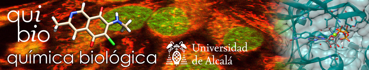
Publicaciones > Marcelo et al
Nonlinear Emission of Quinolizinium Based Dyes With Application in Fluorescence Lifetime Imaging.
1. Centro de Química-Física Molecular (CQFM) and Institute of Nanoscience and Nanotechnology (IN), Instituto Superior Técnico, Universidade de Lisboa, Av. Rovisco Pais, 1, 1049-001 Lisboa, Portugal. 2. Departamento de Química Orgánica y Química Inorganica, Universidad de Alcalá, 28871, Alcalá de Henares, Madrid, Spain.
Abstract
Charged molecules based on the quinolizinum cation have potential applications as labels in fluorescence imaging in biological media under nonlinear excitation. A systematic study of the linear and nonlinear photophysics of derivatives of the quinolizinum cation substituted by either dimethylaniline or methoxyphenyl electron donors is performed. The effects of donor strength, conjugation length, and symmetry in the two-photon emission efficiency are analyzed in detail. The best performing nonlinear fluorophore, with two-photon absorption cross sections of 1140 GM and an emission quantum yield of 0.22, is characterized by a symmetric D-π-A+-π-D architecture based on the methoxyphenyl substituent. Application of this molecule as a fluorescent marker in optical microscopy of living cells revealed that, under favorable conditions, the fluorophore can be localized in the cytoplasmatic compartment of the cell, staining vesicular shape organelles. At higher dye concentrations and longer staining times, the fluorophore can also penetrate into the nucleus. The nonlinearly excited fluorescence lifetime imaging shows that the fluorophore lifetime is sensitive to its location in the different cell compartments. Using fluorescence lifetime microscopy, a multicolor map of the cell is drafted with a single dye.